Radiologists develop point-of-care AI for chest X-rays
SaveSavedRemoved 0
[ad_1]
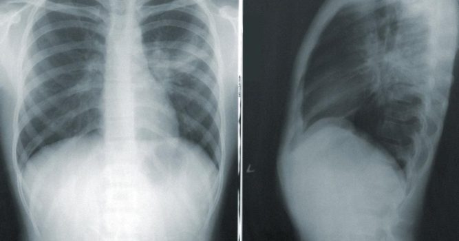
The model’s development involved 3,278 chest radiographs from five different sites. A chest radiologist reviewed the images and categorized the reasons for their suboptimality. The de-identified images were then uploaded into an AI server application for training and testing purposes. Model performance was measured based on its area under the curve (AUC) for differentiating between optimal and suboptimal images.
Reasons for suboptimality were categorized as either missing anatomy, obscured thoracic anatomy, inadequate exposure, low lung volume or patient rotation. For accuracy in…
[ad_2]
Go to publisher site for the complete article:
Read More

